TIL composition and source
Antigen and APC configuration
Recognition and target cell activity
Tumor residence does not establish tumor recognition
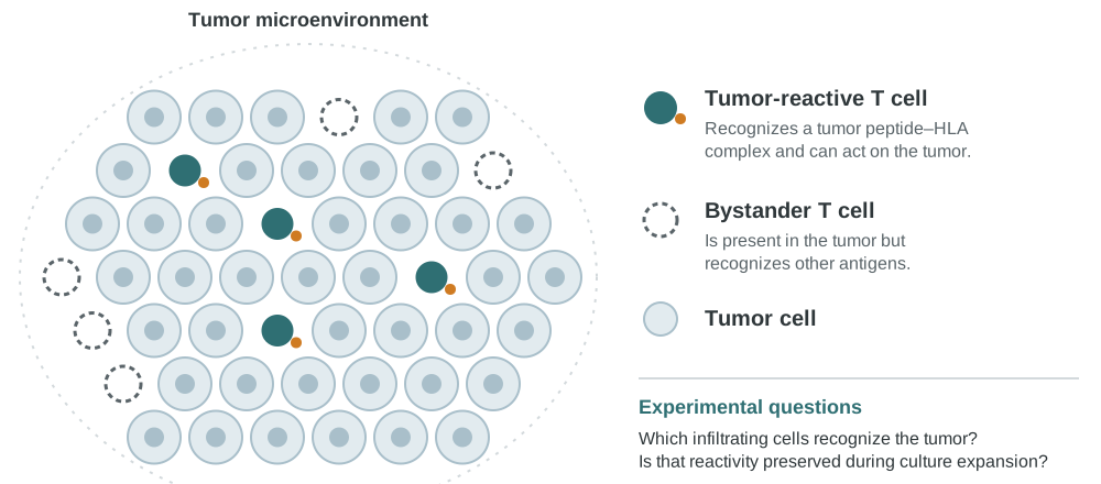
T cells found within a tumor can have different antigen specificities and functional states. Simoni and colleagues documented substantial populations of bystander CD8 T cells in human tumor infiltrates. This finding illustrates why the location of a T cell is not sufficient evidence of tumor reactivity. [1]
For an experimental culture, record the starting population and distinguish the total number of lymphocytes from the number of cells responding to the antigen or target being studied.
Cell abundance and tumor reactivity answer different questions
A tumor sample may contain T cells recognizing tumor antigens, cells responding to unrelated antigens and regulatory populations. Their representation varies by donor, tissue, antigen and treatment history. Tumor reactivity describes a functional property, not a single definitive surface marker.
Compare the starting population with the cultured response using antigen-positive and antigen-negative targets, appropriate HLA controls, viability and target cell activity. If antigen-directed APC stimulation is part of the design, test whether it changes the relevant functional fraction rather than assuming that more cells means more tumor recognition.
Repeated challenge can address persistence of activity in the assay, while cytokine measurements and phenotype provide complementary information.
Use phenotyping to inform a functional test
In a melanoma study, Gros and colleagues found that PD-1 expression identified a tumor-reactive CD8 repertoire, including neoantigen-specific cells, within the tumors examined. The observation shows how phenotype can inform enrichment or investigation in a defined setting. It should not be treated as a universal selection rule for every tumor or culture. [2]
Phenotypic information is most useful when connected to direct recognition assays. A marker-defined population may include functionally different cells, and culture can alter the characteristics measured at the beginning of the experiment.
Assess what changes during culture
Expansion creates enough material for subsequent analysis, but cell yield and biological quality are separate outcomes. A culture can increase total numbers without increasing the fraction that recognizes the intended antigen.
| Measurement | Research question |
|---|---|
| Cell yield & viability | How much viable material is available? |
| Population composition | Which cell populations increased or decreased? |
| Antigen-specific recognition | Did the relevant response persist or change? |
| Target cell function | Does the culture recognize an appropriate tumor target? |
| Culture history | How do stimulation and sampling conditions relate to the result? |
Why the APC configuration matters
An antigen-expressing APC can help organize a study around a specified recognition event. Antigen-directed stimulation could be investigated as a way to alter the responding population, but enrichment and preservation of function must be measured rather than assumed.
Use appropriate antigen and HLA comparisons, define the immune cell source and test a relevant target. When comparing stimulation strategies, keep sampling time, passage context and functional assay conditions comparable.
Planning an antigen-directed eDC study
A renewable antigen-expressing eDC configuration provides a possible APC component for a defined T cell study. Its suitability for a particular TIL population requires project-specific evidence. A custom modification can be considered when it addresses an explicit APC gene function question.
The useful deliverable is an interpretable relationship between the cellular configuration and the measured response. This article does not claim that eDC stimulation improves clinical TIL efficacy, reduces manufacturing costs or preserves a particular phenotype without supporting data.
References
- Simoni Y et al. Bystander CD8+ T cells are abundant and phenotypically distinct in human tumour infiltrates. Nature. 2018;557:575–579. DOI: 10.1038/s41586-018-0130-2.
- Gros A et al. PD-1 identifies the patient-specific CD8+ tumor-reactive repertoire infiltrating human tumors. Journal of Clinical Investigation. 2014;124:2246–2259. DOI: 10.1172/JCI73639.