Renewable, antigen-defined APC systems
The eDC platform provides a cellular foundation for studying antigen-specific immune function and engineering APC configurations around defined research questions.
eDC (engineered dendritic cells) are renewable human antigen-presenting cell lines established from primary blood cells. They keep a mature, activated dendritic cell phenotype in culture, expand to supply repeated experiments, and accept a defined antigen and agreed gene modifications. The eDC system is the basis of the APC drug discovery platform.
The platform brings together antigen-expressing eDC lines, paired target cell assay systems and supporting culture cytokines. Custom development begins with customer-provided PBMCs and may include agreed genetic modifications.
Specific antigen configuration
T cells suited to the study
Matched target and controls
Mature, activated dendritic cell phenotype
Flow cytometry shows a lineage-negative, myeloid dendritic cell profile with antigen presentation, costimulatory and lymph node homing markers. CrossRelay reports that eDCs prime naïve T cells to antigen-specific cytotoxic effector cells; the underlying work is published. [PNAS 2018]
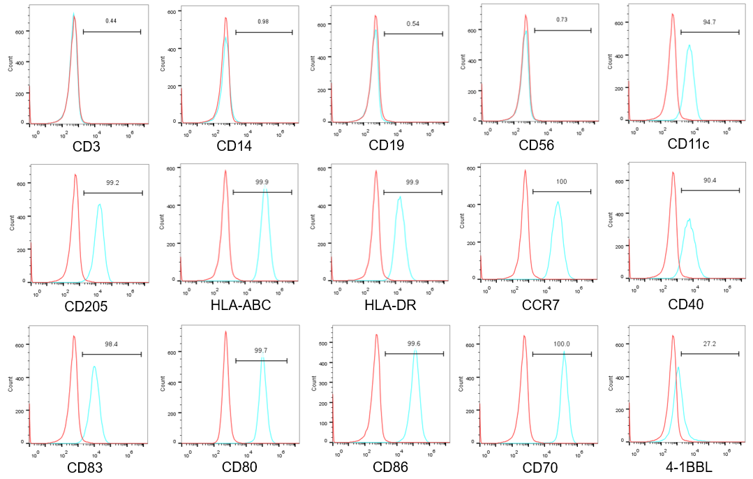
eDC immunophenotype. Single-parameter histograms for 15 markers, with the gate values printed on the panel. Red and cyan traces are shown as in the source figure. Control definition, line identity, passage and gating are not stated on this page, so the panel is descriptive and is not a release specification.
Antigen presentation
HLA-ABC (99.9), HLA-DR (99.9)
Costimulation and activation
CD80 (99.7), CD86 (99.6), CD83 (98.4), CD40 (90.4), CD70 (100.0); 4-1BBL (27.2, modest shift)
Migration
CCR7 (100)
Myeloid dendritic cell identity
CD11c (94.7), CD205 (99.2)
Lineage markers, not shifted
CD3 (0.44), CD14 (0.98), CD19 (0.54), CD56 (0.73)
Values in brackets are the gate percentages printed on the panel. Groups summarize histogram shifts relative to the overlaid trace. The panel does not assign an eDC to a classical DC subset and does not establish priming function, which requires functional assays.
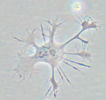
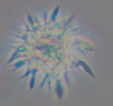
Original phase-contrast micrographs shown at native size. Magnification, scale bars and line identity are not specified. Morphology does not establish antigen-specific function.
eDC technology compared with the conventional MoDC method
MoDCs are the most widely characterized DC format in research. The approved autologous product sipuleucel-T is made from blood mononuclear cells that include antigen-presenting cells, so it is not a pure MoDC preparation. [24] The table lists characteristics reported by CrossRelay for eDC; confirm each parameter on the specific line.
| Feature | Conventional MoDC method | eDC technology |
|---|---|---|
| Starting material | Donor monocytes, often from leukapheresis in a hospital setting | Primary PBMCs; a small blood draw is the described starting sample |
| Growth in culture | Differentiated from monocytes with GM-CSF and IL-4; not propagated | Grows in culture, supplying cells for repeated experiments |
| Maturation and activation | Additional cytokines or TLR ligands are added after differentiation | Mature, activated phenotype maintained in culture |
| Standardization | Donor-dependent yield and quality | One defined line across experiments |
| Genetic modification and antigen loading | Lower efficiency, as reported | Lentiviral transduction, mRNA-LNP, and antigen uptake with cross-presentation |
| Format | Autologous | Autologous, or HLA-matched allogeneic |
| Lymphoid homing | Lower, as reported | CCR7 expressed; homing function requires testing in the intended model |
| Storage | Short-term | Long-term cryostorage; viability above 85% is reported for eDC |
Connect the components of the experiment
| Component | Research role | Explore |
|---|---|---|
| Antigen-expressing eDC | Provides an APC configuration around the study antigen | eDC lines › |
| Paired target cell | Enables functional assessment with antigen/HLA comparisons | Paired systems › |
| Supporting cytokines | Supports connected immune cell culture workflows | Culture support › |
| Custom modifications | Enables project-specific APC gene function questions | Custom services › |
Characterize the selected configuration
For repeated experiments, assess renewal alongside antigen expression and functional activity at the relevant passages. For gene function studies, confirm the requested modification and include matched controls.
Explore representative functional dataMoDCs and progenitor-derived DCs are useful research systems. Compare antigen/HLA matching, culture context and the actual functional endpoint before selecting an eDC configuration.
Read the balanced DC-method comparisonResearch applications
Build the assay around the endpoint: antigen-specific response, target cell function or the effect of a selected APC gene modification.
Explore research applicationsSee how the platform supports target discovery and validation