Defined cells and collection conditions
Identity and co-isolate assessment
Recipient cells and appropriate controls
Begin with accurate terminology
Extracellular vesicles (EVs) are membrane-enclosed particles released by cells. The term exosome describes an endosomal origin, which cannot be established from a small particle size alone. MISEV2023 recommends terminology and characterization appropriate to the evidence available. [1]
This distinction matters when comparing studies. An EV preparation can contain several particle populations, as well as co-isolated material. A shared label does not establish equivalent composition, purity or function.
Antigen presentation beyond the source cell
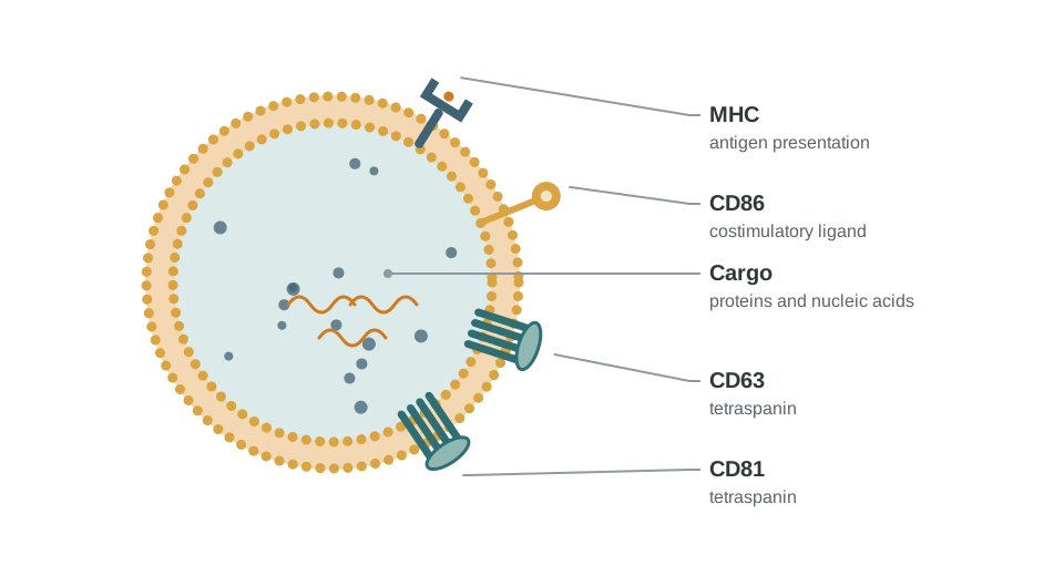
Early work showed that DC-derived vesicles could carry functional MHC molecules and costimulatory components, and that antigen-associated preparations could induce antitumor responses in mouse models. This established a research rationale for examining vesicle-mediated immune communication. [2]
The route by which a preparation influences immune cells depends on the system. Experiments should distinguish direct interactions from effects mediated by recipient APCs or soluble material carried through the isolation process. Activity observed for a particular preparation cannot be assumed for every DC-derived EV sample.
Ask what the vesicle carries and what it does
Membrane-associated HLA, costimulatory molecules and cargo are potential features to characterize in DC-derived preparations. Their presence and abundance depend on the source cell state and isolation method. A schematic cannot establish antigen presentation or activation potency.
Characterize particle abundance and size, membrane-associated markers, non-vesicular contaminants and the relevant antigen or peptide–HLA signal. CD63 or CD81 alone does not establish a preparation’s purity or assign every particle to a specific vesicle subtype.
Then test biological activity with suitable responder cells and controls for soluble factors or co-isolates. Compare antigen-dependent effects with the corresponding negative preparation. These are research questions for immune communication; therapeutic efficacy requires separate evidence.
Separate identity from biological activity
Characterization should connect the source cell state and collection conditions with particle properties, associated proteins and the intended functional endpoint. MISEV2023 provides a framework for documenting these issues and for examining non-vesicular contributions. [1]
| Evidence | Reason to include it |
|---|---|
| Source & collection conditions | Defines the starting biological context |
| Particle characterization | Describes the preparation using complementary methods |
| Co-isolate assessment | Helps distinguish vesicle-associated effects from other material |
| Functional controls | Tests the proposed activity and relevant alternative explanations |
| Repeat preparation | Assesses whether the observation persists across independent collections |
Measuring activity in a biological context
A vesicle-associated antigen or cargo does not establish delivery to the intended tissue, an effective dose or a clinical benefit. These questions require their own evidence. Claims of inherent low toxicity, universal targeting or guaranteed immune activation are therefore too broad for a research resource.
The useful next experiment is specific: identify the recipient cell, define the proposed signal and test whether the measured activity depends on the characterized preparation.
Questions relevant to renewable APC systems
A renewable eDC configuration could provide a defined source cell context for exploratory immune communication research. That is a research question to validate, rather than evidence of an established EV production service.
Comparisons would need to document the antigen-expressing eDC configuration, culture state, vesicle preparation and functional endpoint. CrossRelay’s current product scope remains eDC lines, paired assay systems and supporting cytokines; this article provides broader scientific context.
References
- Welsh JA et al. Minimal information for studies of extracellular vesicles (MISEV2023): From basic to advanced approaches. Journal of Extracellular Vesicles. 2024;13:e12404. DOI: 10.1002/jev2.12404.
- Zitvogel L et al. Eradication of established murine tumors using a novel cell-free vaccine: dendritic cell-derived exosomes. Nature Medicine. 1998;4:594–600. DOI: 10.1038/nm0598-594.